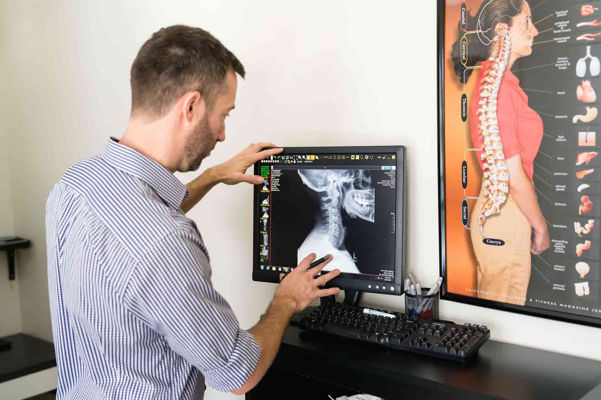
An MRI and Blair upper cervical X-rays answer different questions about your neck. An MRI is excellent at showing soft tissues like discs, nerves, and the spinal cord, which makes it valuable for spotting herniations and other damage. Blair X-rays are taken at angles specific to your anatomy to reveal exactly how your top two vertebrae are positioned. One shows what the tissue looks like, and the other shows precisely how the bones have shifted.
What an MRI Shows
An MRI, or magnetic resonance imaging, uses powerful magnets and radio waves to create detailed images of the soft tissues in your body. For the spine, that means a clear view of your discs, nerves, spinal cord, and surrounding structures.
This makes an MRI a powerful tool for identifying certain problems. A herniated disc, a tumor, nerve compression, or damage to the spinal cord will typically show up clearly on an MRI.
If a doctor suspects soft-tissue damage or needs to rule out something serious, an MRI is often the right choice. It is very good at what it is designed to do, which is show the condition of the tissue.
That strength is also its boundary. An MRI shows what the tissue looks like, but not the exact angular position of the individual vertebrae in your upper neck.
What Blair Upper Cervical X-rays Show
Blair X-rays are a different kind of imaging with a different purpose. Rather than showing soft tissue, they capture the precise position and orientation of your atlas and axis, the top two bones in your neck.
What sets them apart is the approach. Before taking them, we assess the unique angles of your individual anatomy, then position the images to match. That customization is what allows the X-rays to reveal the exact direction and degree of a misalignment.
A standard neck X-ray taken from generic angles cannot show this the same way. The whole point of the Blair series is to map how your specific vertebrae have shifted so a correction can be calculated from real data.
Think of it like the difference between a photo of a room and a set of blueprints. A photo shows you what is there, while blueprints give you exact measurements you can build from. Blair X-rays are closer to blueprints for your upper neck, which is precisely what an accurate correction requires.
MRI vs. Blair X-rays: Different Tools for Different Jobs
Here is how the two compare:
- What they image: An MRI shows soft tissue like discs and nerves. Blair X-rays show the bony position of the atlas and axis.
- Best for: An MRI is best for detecting herniations, tumors, and tissue damage. Blair X-rays are best for measuring upper cervical misalignment.
- How they are taken: An MRI follows a standard protocol. Blair X-rays are angled to each patient’s individual anatomy.
- The question they answer: An MRI asks what the tissue looks like. Blair X-rays ask how the bones are positioned.
Neither is better in a general sense. They are built for different jobs, and the right one depends on what you are trying to find out.
Why Blair Requires Its Own Specialized X-rays
People sometimes ask why a recent MRI is not enough to guide an upper cervical adjustment. The answer comes down to precision and purpose.
An MRI can confirm that a disc is herniated, but it does not tell us the exact angle at which your atlas has rotated or tilted. Correcting the upper neck without that information would be guesswork, and guesswork is the opposite of what this technique is about.
The specialized X-ray series exists to give us a precise map. From that map, we calculate a correction angle specific to your anatomy. You can read more about how this works on our Blair upper cervical chiropractic page.
What Kind of X-rays Does Blair Use?
The Blair series is not a single snapshot. It involves a set of images taken from specific angles that are calculated for your individual anatomy.
This matters because the atlas and axis are shaped a little differently in every person, and their joint surfaces sit at unique angles. Generic views would flatten that detail and hide the very misalignment we are trying to measure.
By tailoring the angles to you, the images show the true position of these vertebrae. That is what allows a correction to be planned with precision rather than estimated.
Do the Two Ever Work Together?
They can, and sometimes they complement each other well. If you arrive with a known disc problem, an MRI may already have identified it, which helps us understand your full picture.
The Blair X-rays then add the piece the MRI was never meant to provide: the precise positioning of your upper cervical spine. Someone dealing with a herniated or bulging disc alongside upper cervical symptoms might benefit from what both types of imaging offer.
We see this combination regularly at our Austin practice. The point is not to pit one against the other, but to use the right tool for the right question.
What About Radiation From X-rays?
It is a fair question, and one patients often raise. X-rays do involve a small amount of radiation, while an MRI uses none.
The exposure from a Blair series is low, and it is weighed carefully against the value of the information it provides. Correcting your upper neck accurately requires seeing exactly how it is positioned, and the specialized X-rays are what make that possible.
We take only the images needed to map your anatomy, nothing extra. The aim is always the most precise correction with the least imaging required.
Does an MRI Ever Miss an Upper Cervical Problem?
An MRI does not exactly miss the upper neck, but it is not built to analyze it the way Blair imaging does. An MRI can show that the discs and soft tissue look healthy while an alignment problem still exists.
The reason is that an MRI captures structure, not the precise positional relationships between the atlas, axis, and skull. Those relationships are what define an upper cervical misalignment.
So a clear MRI is genuinely good news about your soft tissue, but it does not rule out the kind of misalignment that a specialized upper cervical analysis is designed to find.
What This Means for You as a Patient
If you have had an MRI and still do not have answers about ongoing neck-related symptoms, that does not mean the MRI failed. It may simply mean the issue lies in something the MRI was not designed to measure.
An upper cervical evaluation looks at your neck from a different angle, quite literally. For patients in Austin who have felt stuck after inconclusive imaging, it can be the missing piece. You can learn what a first visit involves on our new patients page.
None of this is about choosing sides between two types of imaging. It is about understanding that a clear scan of one kind does not always mean every question has been answered. Sometimes the next step is simply a different way of looking.
If you want a clearer picture of what is happening in your upper neck, schedule a visit with Full Life Chiropractic in Austin or call 512-953-9612.